Skeletal Class III

Orthognathic surgery Mandible forward

Skeletal Class III malocclusion surgery is an orthognathic surgery procedure that corrects skeletal abnormalities that cause a misalignment between the upper and lower teeth. Class III is characterized by excessive overhang of the lower teeth relative to the upper teeth due to abnormal positioning of the lower jaw (mandible) relative to the lower jaw (maxilla).

What is a skeletal Class III malocclusion?

woman with chin forward

In the case of severe skeletal Class III , orthodontic correction at the dentist with braces will not be sufficient. Surgery for skeletal Class III corrects imbalances in the jaw bones.

Assessment

The process usually begins with a thorough evaluation by the orthodontist who begins work and then refers the patient to a maxillofacial surgeon . X-rays and dental impressions are taken to assess the nature and extent of the malocclusion (bad correspondence between the teeth). Then, surgery may be performed on the maxilla and/or mandible.

Operation

During surgery, precise cuts are made to the jaw bones to allow their repositioning. Once orthognathic surgery is performed, postoperative orthodontic treatment may be necessary to adjust the position of the teeth.

Skeletal Class III surgery is reserved for patients with a significant misalignment between the jaws. The treatment process is lengthy and requires close collaboration between the orthodontist and the maxillofacial surgeon or oral surgeon to ensure the success of the operation.

forward jaw prognathism class III surgery
Diagram of bimaxillary orthognathic surgery for a skeletal Class III malocclusion.

Is Class III skeletal surgery reimbursed?

The management of orthognathic surgery for the correction of a Class III malocclusion depends on the severity of the malocclusion and the functional consequences.

Health Insurance (Social Security): Social Security may cover part of the costs associated with orthognathic surgery if it is considered medically necessary (functional disorders). However, Social Security does not cover all costs, and costs may remain.

Mutual insurance: Supplemental health insurance plays an important role in covering remaining costs. You should contact your mutual insurance company to find out the reimbursement terms specific to your policy.

Before undergoing any surgical procedure, it is recommended that you consult your maxillofacial surgeon, orthodontist, and insurance company for specific coverage information. This will help you understand any approval procedures and the costs you will be responsible for.

Why perform skeletal class III surgery?

Aesthetic improvement

A Class III malocclusion means an unbalanced facial profile, excessively protruding lower teeth, or a lower jaw that is too far forward. Orthognathic surgery improves facial appearance by restoring harmony and facial proportions.

Functional problems

A Class III malocclusion can cause functional problems such as difficulty chewing, speaking, or muscle tension. Surgery can correct these problems by realigning the jaws to promote proper occlusion (mouth closure). The lower incisors often protrude in front of the upper ones.

Before/After Simulations of Class III Skeletal Surgery

surgery chin before after 800

Is surgery for a skeletal Class III painful?

pain surgery

Pain during surgery

The operation is performed under general anesthesia. Therefore, the patient is unconscious during the procedure and feels no pain. As a result, the procedure itself is painless.

Postoperative pain

After surgery, it is normal to have some pain and feel pressure and discomfort. However, most patients manage their pain well with pain relief medications prescribed by the surgeon and a cryotherapy band.

Swelling and bruising

Swelling and bruising in the lower face are common. While not painful, it can cause discomfort. Topical cold and anti-inflammatory medications can help reduce swelling.

Respiratory discomfort

Temporary breathing discomfort may occur during the recovery period.

Sensitivity to touch

The operated area is sensitive to touch for a while. This sensitivity decreases as healing progresses.

How to find a surgeon for a skeletal class III?

surgery

Recommandations

Discuss it with your loved ones. Their experiences and recommendations can be invaluable.

Reviews

Look for online reviews of surgeons. However, be critical and consider the diversity of experiences. Don’t rely solely on reviews.

Initial consultation

Schedule consultations with multiple surgeons to discuss your goals, show them your before/after simulation, and see examples of surgical results.

What are the risks of surgery (skeletal class III)?

1. Bleeding
Although bleeding is usually controlled during surgery, there may be a risk of excessive bleeding postoperatively.

2. Infections
Infections are always a risk after surgery. Surgeons take precautions to minimize this risk, but it can still occur.

3. Anesthetic Reactions
As with any surgical procedure, there is a risk of allergies, adverse reactions or other complications related to anesthesia.

4. Bruising and Swelling
Bruising (accumulation of blood) and swelling are common side effects after surgery. They can contribute to postoperative discomfort and take time to resolve.

5. Bone problems
Orthognathic surgery involves structural changes to the jaw bones. There is a risk of poor bone healing.

6. Disorders
During the recovery period, you may have temporary difficulty eating or speaking.

7. Temporomandibular Joint (TMJ) Problems
Joint problems, although rare, can occur after surgery and affect the function of the temporomandibular joints.

8. Aesthetic dissatisfaction
Although the goal of surgery is to improve facial aesthetics, there is a risk of post-operative disappointment.

What are the steps involved in an operation to correct a protruding mandible?

1) Initial consultation

The process begins with an initial consultation with an orthodontist . During this phase, x-ray images, dental impressions, and facial photographs may be taken to assess the severity of the malocclusion. Once the need for orthognathic surgery is established, the patient is referred to a maxillofacial surgeon.

2) Simulation

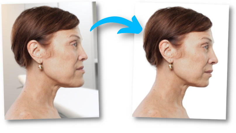
A simulation can be performed to simulate the outcome and help the patient visualize the desired result. The before/after simulation also allows you to clearly explain to your surgeon what you want from the operation. Perform a simulation of a Class III skeletal surgery.

The maxillofacial surgeon will conduct a thorough evaluation, including clinical examinations, medical imaging, and a detailed analysis of the malocclusion. A precise surgical plan is developed to determine the necessary jaw movements.

3) Preparation for the intervention

Before surgery, the patient often undergoes preoperative orthodontic treatment to align the teeth and prepare the jaws for surgery. This involves wearing braces for several months or years.

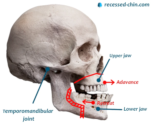
4) Day of intervention – Skeletal Class III

During surgery, the maxillofacial surgeon makes precise cuts in the jawbones to reposition them. The bones are secured with titanium screws and plates. They are biocompatible and do not beep at airports.

5) Post-operative period

After surgery, the patient is usually monitored for one day in the hospital. The length of hospitalization depends on the complexity of the procedure and the patient’s recovery: 1 to 3 days.

The patient then continues to recover at home, following the maxillofacial surgeon’s instructions. This involves a liquid or soft diet for a period of time and taking precautions to avoid stress on the recently operated jaws.

6) Follow-up

After the operation, the patient continues orthodontic treatment to adjust the position of the teeth and perfect the occlusion.

Developments after orthognathic surgery

End of operation

The first few days after surgery are spent in the hospital for postoperative monitoring. Care includes pain management, bleeding control, and assessing the patient’s general condition.

1st week

The diet is liquid to avoid any stress on recently operated jaws.

1st month

Rest is essential during this phase. Physical activities are limited, and the patient must follow the maxillofacial surgeon’s instructions for postoperative care, including cleaning scars and teeth.

After several months

Postoperative orthodontic treatment begins to adjust the position of the teeth.

Speech therapy

Then, physical rehabilitation or speech therapy sessions are recommended to facilitate recovery and restore normal function of the jaws and facial muscles.

Follow up

Regular follow-up visits with the maxillofacial surgeon are scheduled to monitor healing, adjust medications as needed, and address any patient concerns.

face surgery jaw
face surgery jaw
face surgery jaw
face surgery jaw

What are the benefits of ultrasonic cutting surgery?

This technique has advantages in maxillofacial and dental surgery.

Less damage to soft tissue

Piezoelectric instruments are specially designed to target bone tissue while sparing surrounding soft tissue, such as nerves, blood vessels and mucous membranes.

Less bleeding

The ultrasonic cutting technique causes less bleeding than traditional methods, improving the surgeon’s visibility during the procedure.

Less trauma

Piezo surgery reduces overall tissue trauma, resulting in faster post-operative recovery and less pain for the patient.

Fewer complications

Due to its precision, piezo surgery helps reduce the risk of complications such as unintentional fractures or nerve damage.

Diversified application

Piezoelectric instruments are used for tooth extraction, site preparation for dental implants, periodontal surgery and orthognathic surgery.

Less vibration

Unlike other cutting methods, piezo surgery generates less vibration, which is beneficial in sensitive procedures.

Improved postoperative recovery

Many patients report faster post-operative recovery and reduced discomfort compared to traditional surgical methods.

What scars are left by orthognathic surgery?

The incisions for jaw and maxilla surgery are made inside the mouth. The surgeon places stitches inside the mouth with absorbable thread. There are no visible scars from the outside.

History of orthognathic surgery

Beginnings of maxillofacial surgery (19th century)

The first surgeries on the maxillofacial bones were performed in the 19th century. However, at that time, understanding of facial growth and bone relationships was limited, and results were often unpredictable.

Early orthognathic surgery

During the first half of the 20th century, orthognathic surgery made significant progress thanks to the work of pioneering surgeons. They contributed to the understanding of jaw dysfunctions and the development of surgical techniques to correct them.

Evolution of surgical planning

The 1950s and 1960s saw the introduction of new surgical planning methods, including the use of plaster models and cephalometric radiographs. These advances allowed for better anticipation of necessary jaw movements.

Development of internal fixation surgery

The use of titanium plates and screws to hold bones in position revolutionized orthognathic surgery in the 1970s. This allowed for increased stability of surgical results.

Use of computer techniques

With the advent of computers and advanced medical imaging, surgeons can use computer-aided planning techniques to precisely design the necessary surgical movements.

Minimally invasive surgery

Orthognathic surgery techniques have continued to evolve toward more minimal and less invasive approaches, thereby reducing visible scarring and speeding recovery.

Integration of orthodontics

Today, orthognathic surgery is often integrated into comprehensive orthodontic treatment plans. Preoperative orthodontics is used to prepare the teeth and facilitate surgery. Postoperative orthodontics is necessary to adjust the final position of the teeth.

Before/After Class III Skeletal Simulations

surgery simulation
Shopping Cart
Scroll to Top